Posterior instrument的問題,透過圖書和論文來找解法和答案更準確安心。 我們找到下列包括價格和評價等資訊懶人包
Posterior instrument的問題,我們搜遍了碩博士論文和台灣出版的書籍,推薦林茂雄寫的 牙材力:大師們的百寶箱 和Agarwal, Amar (EDT)的 More Phaco Nightmares: Conquering Cataract Catastrophes都 可以從中找到所需的評價。
另外網站Posterior spinal instrumentation and decompression with ...也說明:Posterior lumbar instrumentation requires sufficient primary stiffness to ensure bony fusion and to avoid pseudarthrosis, screw loosening, or implant failure.
這兩本書分別來自林茂雄 和所出版 。
國立陽明交通大學 教育研究所 段正仁所指導 蔡尚恆的 以fMRI探討不同推理能力之理科生在光學透鏡成像操弄時之心智旋轉腦神經作用機轉 (2021),提出Posterior instrument關鍵因素是什麼,來自於功能性磁振造影、心智模型、心智旋轉、空間關係性推理、關係整合能力、光學透鏡成像操弄、關係性推理能力。
而第二篇論文國立陽明交通大學 光電工程研究所 孫家偉所指導 蕭天語的 智慧瓊斯矩陣光學同調斷層掃描術之開發與應用 (2021),提出因為有 瓊斯矩陣光學同調斷層掃描術、穆勒矩陣、深度學習、牙結石、淋巴瘤、膠質瘤、神經纖維的重點而找出了 Posterior instrument的解答。
最後網站Posterior instrumentation with release - AO Surgery Reference則補充:Most cases can be managed with a posterior osteotomy and pedicle screw instrumentation. The main indications for anterior release is advanced disk degeneration ...
除了Posterior instrument,大家也想知道這些:
牙材力:大師們的百寶箱
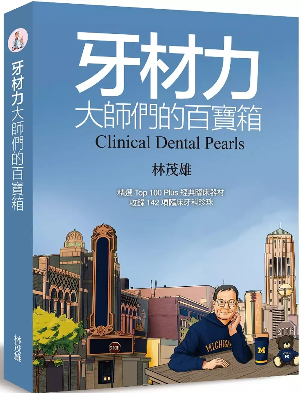
為了解決Posterior instrument 的問題,作者林茂雄 這樣論述:
Top 100 Plus 經典臨床牙科器材,142項臨床牙科珍珠;牙醫師、牙技師與牙材商溝通的橋梁。 ◎《牙材力:大師們的百寶箱》就是你的超能力── ● 濃縮數千篇文獻的精華,快速提升你的《牙材力》 ● 牙醫學生、牙醫師、牙材廠商,每人必備牙材手冊 ● 牙科材料超速學習,一次搞懂牙材分類、選擇標準及臨床使用 ● 142 項牙科珍珠產品優缺點、臨床應用時機,與使用訣竅 ● 牙醫師、牙技師與牙材廠商共同的語彙、溝通的橋梁 材料學在牙醫科學研究範疇內更見其精髓,任何一項新產品的推出,都是一項挑戰!牙醫界近幾年
的突飛猛進,更容易考驗這項說法! 《牙材力:大師們的百寶箱》精選Top 100 Plus 經典臨床器材,根據分類順序排列方式,一一介紹每個產品的特點、臨床應用和操作訣竅,是學生的基本修煉,醫師的臨床寶鑑。
以fMRI探討不同推理能力之理科生在光學透鏡成像操弄時之心智旋轉腦神經作用機轉
為了解決Posterior instrument 的問題,作者蔡尚恆 這樣論述:
本研究補充了過去對光學空間關係性推理所顯露的心智旋轉機制文獻極少的問題,亦解決了相關主題的腦波文獻空間解析度相對不足的限制。本研究目的在探討不同關係性推理能力的理科生,於不同關係整合能力需求程度的光學成像推理任務中,其心智旋轉的歷程,大腦活化的腦區與活化程度在fMRI動態變化之探究。本研究包含「fMRI光學成像推理實驗」,受試者會在反射屏幕回答三類光學關係性推理問題(單一凹透鏡、單一平面鏡、單一凸透鏡);之後會有「瑞文氏測驗」測其「關係性推理能力」。研究對象為20歲到30歲受過理工教育之自願者18名。資料蒐集工具為在Matlab運作下的cogent,它紀錄了行為資料「成像結果作答正確率、作答
反應時間」,cogent也呈現刺激材料和實驗時序排程;fMRI紀錄了「不同腦區的BOLD signal活化強度、活化位置、活化範圍」;瑞文氏測驗紙紀錄「答題正確率」。研究結果顯示三類光學成像推理任務之作答反應時間、正確率有差異,其行為資料結果顯示:推理任務難度凸透鏡>平面鏡>凹透鏡。瑞文氏分數與三類光學成像推理任務之作答反應時間、正確率有相關,其線性迴歸結果顯示:關係性推理能力可預測最困難任務之「凸透鏡成像結果作答正確率」;作答反應時間與正確率呈負相關。此外三類光學成像推理任務在「心智模型操弄推理階段」大致牽涉「BA19、PPC(BA7、39、40)、BA4&6、IFG、DLPFC(BA9、4
6)、BA10&32」等腦區的活化,其fMRI資料的結果顯示,於難度最高的「凸透鏡心智模型操弄推理階段」所活化腦區最廣、活化強度最高,主要涵蓋腦部頂葉連結到BA6與心智旋轉密切關聯之神經網路。更發現「平面鏡心智模型操弄推理階段」比「凸透鏡心智模型操弄推理階段」可能更多著重腹側視覺訊息流(ventral visual stream)處理物體辨識圖像之訊息。此外在「心智模型操弄推理階段」任務難度會影響「BA19、PPC(BA7、39、40)、BA4&6、IFG、DLPFC(BA9、46)、BA10&32」等腦區的活化,其fMRI資料的結果顯示,於「凸透鏡對比於凹透鏡的心智模型操弄推理階段」時,活化
有顯著差異的腦區更涵蓋小腦、基底核與Insula。基底核與心智旋轉之關聯可能在此深層神經結構與前額葉及頂葉有神經網路的連結。Insula可能與心智模型形成時不確定性的抉擇有關,這可說明為何凸透鏡成像結果作答正確率最低。此外瑞文氏分數與三類光學成像推理任務在「心智模型操弄推理階段」所活化腦區有部分相關,其線性相關結果顯示,關係性推理能力與「凸透鏡對比於平面鏡的心智模型操弄推理階段」於ACC、Cingulate Gyrus呈正相關,此部分活化的腦區是靠中後方的ACC,此區域與BA6及prefrontal cortex可能有緊密的神經連結,此結果意涵在任務難度差異大的情境下,關係性推理能力越高則「專
注、偵錯、抑制和工作記憶無關訊息、心智旋轉的能力」可能也越強。本研究依任務難度之不同,心智旋轉成像腦區活化型態亦不同且其誘發之活化反應形式相當恆定。此任務可供往後研究心智旋轉腦部運作機制一個好用的工具。本研究釐清部分心智旋轉之腦部運作機制並發現理科生之關係性推理能力與不同難度任務測試下活化腦區之相關性。相信這些成果可為往後探究此領域之人提供重要參酌。(註: 「推理能力」eductive ability,在本論文意同「關係性推理能力」)
More Phaco Nightmares: Conquering Cataract Catastrophes
A PHP Error was encountered
Severity: Warning
Message: file_put_contents(/var/www/html/prints/public/images/books_new/F01/396/62/F013962574.jpg): failed to open stream: Permission denied
Filename: helpers/global_helper.php
Line Number: 140
Backtrace:
File: /var/www/html/prints/application/helpers/global_helper.php
Line: 140
Function: file_put_contents
File: /var/www/html/prints/application/views/article_v2.php
Line: 248
Function: coverWebp_online
File: /var/www/html/prints/application/controllers/Pages.php
Line: 662
Function: view
File: /var/www/html/prints/public/index.php
Line: 319
Function: require_once
A PHP Error was encountered
Severity: Warning
Message: getimagesize(/var/www/html/prints/public/images/books_new/F01/396/62/F013962574.jpg): failed to open stream: No such file or directory
Filename: helpers/global_helper.php
Line Number: 62
Backtrace:
File: /var/www/html/prints/application/helpers/global_helper.php
Line: 62
Function: getimagesize
File: /var/www/html/prints/application/helpers/global_helper.php
Line: 142
Function: coverWebp
File: /var/www/html/prints/application/views/article_v2.php
Line: 248
Function: coverWebp_online
File: /var/www/html/prints/application/controllers/Pages.php
Line: 662
Function: view
File: /var/www/html/prints/public/index.php
Line: 319
Function: require_once
A PHP Error was encountered
Severity: Notice
Message: Trying to access array offset on value of type bool
Filename: helpers/global_helper.php
Line Number: 64
Backtrace:
File: /var/www/html/prints/application/helpers/global_helper.php
Line: 64
Function: _error_handler
File: /var/www/html/prints/application/helpers/global_helper.php
Line: 142
Function: coverWebp
File: /var/www/html/prints/application/views/article_v2.php
Line: 248
Function: coverWebp_online
File: /var/www/html/prints/application/controllers/Pages.php
Line: 662
Function: view
File: /var/www/html/prints/public/index.php
Line: 319
Function: require_once
A PHP Error was encountered
Severity: Notice
Message: Trying to access array offset on value of type bool
Filename: helpers/global_helper.php
Line Number: 66
Backtrace:
File: /var/www/html/prints/application/helpers/global_helper.php
Line: 66
Function: _error_handler
File: /var/www/html/prints/application/helpers/global_helper.php
Line: 142
Function: coverWebp
File: /var/www/html/prints/application/views/article_v2.php
Line: 248
Function: coverWebp_online
File: /var/www/html/prints/application/controllers/Pages.php
Line: 662
Function: view
File: /var/www/html/prints/public/index.php
Line: 319
Function: require_once
A PHP Error was encountered
Severity: Notice
Message: Trying to access array offset on value of type bool
Filename: helpers/global_helper.php
Line Number: 68
Backtrace:
File: /var/www/html/prints/application/helpers/global_helper.php
Line: 68
Function: _error_handler
File: /var/www/html/prints/application/helpers/global_helper.php
Line: 142
Function: coverWebp
File: /var/www/html/prints/application/views/article_v2.php
Line: 248
Function: coverWebp_online
File: /var/www/html/prints/application/controllers/Pages.php
Line: 662
Function: view
File: /var/www/html/prints/public/index.php
Line: 319
Function: require_once

為了解決Posterior instrument 的問題,作者Agarwal, Amar (EDT) 這樣論述:
Even the most experienced cataract surgeon can encounter stressful situations in the operating room. With the new edition of More Phaco Nightmares: Conquering Cataract Catastrophes, surgeons can be prepared to manage unavoidable complications with ease. Dr. Amar Agarwal, along with over 35 of today'
s cataract surgery leaders, explain all there is to know about phacoemulsification and bring their extensive experience with their own surgical nightmares. The book contains 5 sections that gradually escalate from the basics to nightmares, furnishing phaco surgeons with complicated scenarios and the
essential guidance to assess, manage, and resolve. Featuring updated content and brand new, state-of-the-art chapters on a variety of complex situations, More Phaco Nightmares is the toolkit surgeons need to stay in control when facing unique and especially challenging intra- and postoperative comp
lications.Sample chapters include: The Phaco MachineAir Pump, Gas-Forced Infusion, and Active FluidicsIntraoperative Floppy Iris SyndromeSingle-Pass Four-Throw PupilloplastyManagement of Capsule Rupture at Cataract SurgeryMalpositioned Intraocular LensOptic CaptureGlued Intraocular LensManagement an
d Prevention of Negative DysphotopsiaPseudophakic Cystoid Macular Edema More than 350 illustrations and photographs supplement the text, providing visual as well as textual references for each case. Plus, an accompanying video website contains over 4 hours of new, original, high-quality video conten
t, offering additional visual learning to demonstrate the techniques discussed. Offering cutting-edge information, More Phaco Nightmares: Conquering Cataract Catastrophes will guide surgeons on how to mitigate the common problems and unanticipated disasters that may arise for even the most experienc
ed surgeons. Amar Agarwal, MS, FRCS, FRCOphth, is the pioneer of phakonit, which is phaco with needle incision technology. This technique became popularized as bimanual phaco, microincision cataract surgery (MICS), or microphaco. He was the first to remove cataracts through a 0.7-mm tip with the
technique called microphakonit. He also discovered no-anesthesia cataract surgery and FAVIT, a new technique to remove dropped nuclei. This technique is now called sleeveless phaco tip-assisted levitation (SPAL). The air pump, which was a simple idea of using a fish aquarium pump to increase the flu
id into the eye in bimanual phaco and co-axial phaco, has helped prevent surge. This built the basis of various techniques of forced infusion like active fluidics for small incision cataract surgery. He also discovered a new refractive error called aberropia. He was also the first to do a combined s
urgery of microphakonit (700-micron cataract surgery) with a 25-gauge vitrectomy in the same patient, thus having the smallest incisions possible for cataract and vitrectomy. He was the first surgeon to implant a new mirror telescopic IOL (Lipshitz macular implant) for patients suffering from age-re
lated macular degeneration. He was the first in the world to implant a glued IOL. In this, a PC-IOL is fixed in an eye without any capsules using fibrin glue. The Malyugin ring for small pupil cataract surgery was also modified by him as the Agarwal modification of the Malyugin ring for miotic pupil
cataract surgery with posterior capsular defects. Dr. Agarwal’s Eye Hospital also did the first anterior segment transplantation in a 4-month-old child with anterior staphyloma. Dr. Agarwal has brought out the technique of IOL scaffold, in which a 3-piece IOL is injected into an eye between the iri
s and the nucleus to prevent the nucleus from falling down in posterior capsule ruptures. He has combined glued IOL and IOL scaffold in cases of posterior capsule rupture where there is no iris or capsular support and termed the technique glued IOL scaffold. Dr. Agarwal’s Eye Hospital also did a glu
ed endocapsular ring in cases of subluxated cataract for the first time. Pre-Descemet’s endothelial keratoplasty (PDEK) was initiated by Dr. Agarwal. In this, the pre-Descemet’s layer and the Descemet’s membrane with endothelium are transplanted en bloc in patients with diseased endothelium. Dr. Aga
rwal’s Eye Hospital first did contact lens-assisted collagen cross-linking (CACXL), a new technique for cross-linking thin corneas, and they have also worked on E-PDEK, in which an endoilluminator is used to assist in PDEK surgeries. Dr. Agarwal designed the new instrument called the trocar anterior
chamber maintainer, now in complicated cases, which helps give infusion through the anterior chamber and works like a trocar cannula. He also started a new technique of iris suturing called single-pass four-throw (SFT) pupilloplasty. This is used for closed-angle glaucoma and for mydriatic cases. H
e was the principal investigator for the Bausch & Lomb study in the first human eyes using hypersonic vitrectomy with Vitesse. The first 20 surgeries of posterior vitrectomy were done at Dr. Agarwal’s Eye Hospital. He was also the first in the world to use hypersonic vitrectomy with the Vitesse for
a case of posterior capsule rupture with nuclear fragments. He did an anterior vitrectomy, posterior vitrectomy, and nuclear fragment removal with the same Vitesse 23-gauge probe. Dr. Agarwal has performed more than 150 live surgeries at various conferences. His videos have won many awards at the fi
lm festivals of the American Society of Cataract and Refractive Surgery, American Academy of Ophthalmology, and European Society of Cataract and Refractive Surgeons. He has also written more than 75 books, which have been published in various languages (English, Spanish, and Polish). In his center,
he trains doctors from all over the world on phaco, glued IOL, LASIK, and retinal surgeries. He is the chairman and managing director of Dr. Agarwal’s Group of Eye Hospitals, which has 75 eye hospitals across 14 countries all over the world. He can be contacted at [email protected]. The website of
the hospital is http: //www.dragarwal.com.
智慧瓊斯矩陣光學同調斷層掃描術之開發與應用
為了解決Posterior instrument 的問題,作者蕭天語 這樣論述:
中文摘要 ...................................................................... iAbstract ...................................................................... iiAcknowledgement ............................................................... iiiTable of Contents ....................................
......................... ivList of Figures ............................................................... viiList of Tables ................................................................ xList of Abbreviations ......................................................... xi1. Introduction ......
......................................................... 1 1.1 Brief Review of Optical Coherence Tomography (OCT) ...................... 1 1.2 Jones Matrix Optical Coherence Tomography (JM-OCT) ...................... 5 1.2.1 Hardware Designs of Jones Matrix Measurement ......................
.. 5 1.2.2 Algorithms for Jones Matrix Analysis ................................ 7 1.2.3 Fields of Application ............................................... 10 1.3 Deep Learning ........................................................... 11 1.4 Motivation ..................................
............................ 13 1.5 Objective ............................................................... 13 1.6 Organization of this Dissertation ....................................... 142. Principle .................................................................. 16 2.1 Image Formation
of OCT .................................................. 16 2.1.1 Imaging Principle and Selection of Interferometers .................. 16 2.1.2 Chromatic Dispersion Compensation ................................... 19 2.1.3 System and Image Attributes .........................................
20 2.2 Representation and Calculation of Polarization States ................... 21 2.2.1 Overview of Methodologies ........................................... 21 2.2.2 Jones Calculus ...................................................... 24 2.2.3 Mueller Calculus ...........................
......................... 25 2.3 Deep Learning ........................................................... 27 2.3.1 Artificial Neural Network ........................................... 27 2.3.2 Evaluation Metrics .................................................. 29 2.3.3 Model Visualization
Tools ........................................... 313. System Design .............................................................. 35 3.1 Minimalistic Fiber-Optic based JM-OCT ................................... 35 3.1.1 Hardware Setup ...................................................... 35
3.1.2 Software Programming for the Instrument ............................. 43 3.1.3 Optical Formalism ................................................... 46 3.2 Data Processing based on the TensorFlow Framework ....................... 51 3.2.1 Dispersion Compensation ........................
..................... 51 3.2.2 Diagonalization Method .............................................. 54 3.2.3 Concurrent Decomposition Method ..................................... 57 3.3 Training Scheme for Deep Learning ....................................... 62 3.3.1 OCT Data Preparation ..
.............................................. 62 3.3.2 Imaging Preprocessing ............................................... 63 3.3.3 Model Training ...................................................... 664. Clinical Applications ...................................................... 68 4.1
Subgingival Dental Calculus Identification .............................. 68 4.1.1 Model Design and Disease Activation Maps ............................ 70 4.1.2 Performance of Computer-Aided-Detection ............................. 72 4.1.3 Preliminary Result of Dental Calculus using Polarizat
ion Imaging .... 77 4.2 Brain Tumor Classification and Peritumoral Nerve Fiber Visualization .... 80 4.2.1 Classification based on Attention ResNet Model ...................... 81 4.2.2 Brain Tumor Grading based on Transfer Learning ...................... 87 4.2.3 Nerve Fiber Visualization at
Tumoral Boundary ....................... 895. Discussions ................................................................ 95 5.1 Instrument Performance .................................................. 95 5.2 Limitations of the Algorithm ............................................ 96 5.3 Int
egration of TensorFlow-based Frameworks .............................. 97 5.4 Clinical Applications ................................................... 996. Conclusions ................................................................ 101References ..................................................
.................. 104Appendix A Improvements of the Concurrent Decomposition Method ................ 121Appendix B Recruitment of the Brain Tumor Patients and the Data Preparation ... 126